
406M-1
MUC4 (8G7) Mouse Monoclonal Antibody
About This Item
Recommended Products


biological source
mouse
conjugate
unconjugated
antibody form
culture supernatant
antibody product type
primary antibodies
clone
8G7, monoclonal
description
For In Vitro Diagnostic Use in Select Regions (See Chart)
form
buffered aqueous solution
species reactivity
human
packaging
vial of 0.1 mL concentrate (406M-14)
vial of 0.5 mL concentrate (406M-15)
bottle of 1.0 mL predilute (406M-17)
vial of 1.0 mL concentrate (406M-16)
bottle of 7.0 mL predilute (406M-18)
Related Categories
1 of 4
This Item | MABT395 | WH0004585M7 | 290M-1 |
|---|---|---|---|
| biological source mouse | biological source mouse | biological source mouse | biological source mouse |
| conjugate unconjugated | conjugate - | conjugate unconjugated | conjugate unconjugated |
| species reactivity human | species reactivity human | species reactivity human | species reactivity human |
| clone 8G7, monoclonal | clone 8G7, monoclonal | clone 5B12, monoclonal | clone MRQ-17, monoclonal |
| antibody form culture supernatant | antibody form purified immunoglobulin | antibody form purified immunoglobulin | antibody form culture supernatant |
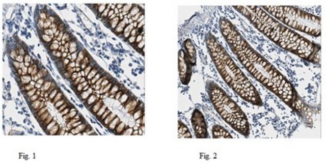
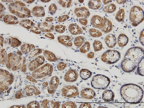
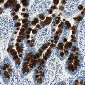
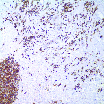
| technique(s) immunohistochemistry (formalin-fixed, paraffin-embedded sections): 1:10-1:50 | technique(s) immunohistochemistry: suitable, western blot: suitable | technique(s) immunohistochemistry (formalin-fixed, paraffin-embedded sections): suitable, indirect ELISA: suitable, western blot: 1-5 μg/mL | technique(s) immunohistochemistry (formalin-fixed, paraffin-embedded sections): 1:50-1:200 |

General description
Quality
 IVD |  IVD |  IVD |  RUO |
Linkage
Physical form
Preparation Note
Other Notes
Legal Information
Not finding the right product?
Try our Product Selector Tool.
Choose from one of the most recent versions:
Certificates of Analysis (COA)
Don't see the Right Version?
If you require a particular version, you can look up a specific certificate by the Lot or Batch number.
Already Own This Product?
Find documentation for the products that you have recently purchased in the Document Library.
Customers Also Viewed
Our team of scientists has experience in all areas of research including Life Science, Material Science, Chemical Synthesis, Chromatography, Analytical and many others.
Contact Technical Service