Skip to Content
MilliporeSigma

Skip To

HPA018304

Sigma-Aldrich

Anti-G3BP2 antibody produced in rabbit

enhanced validation

affinity isolated antibody, buffered aqueous glycerol solution

Synonym(s):

G3BP2 Antibody - Anti-G3BP2 antibody produced in rabbit, G3Bp2 Antibody, Anti-G3BP-2, Anti-GAP SH3 domain-binding protein 2, Anti-Ras GTPase-activating protein-binding protein 2

Sign Into View Organizational & Contract Pricing

About This Item

UNSPSC Code:
12352203
Human Protein Atlas Number:
NACRES:
NA.41
Pricing and availability is not currently available.
Technical Service
Need help? Our team of experienced scientists is here for you.
Let Us Assist

biological source

rabbit

Quality Level

antibody form

affinity isolated antibody

antibody product type

primary antibodies

clone

polyclonal

form

buffered aqueous glycerol solution

species reactivity

human

enhanced validation

orthogonal RNAseq
independent
Learn more about Antibody Enhanced Validation

technique(s)

immunofluorescence: 0.25-2 μg/mL
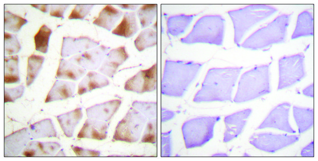
immunohistochemistry: 1:500-1:1000

immunogen sequence

HNDMFRYEDEVFGDSEPELDEESEDEVEEEQEERQPSPEPVQENANSGYYEAHPVTNGIEEPLEESSHEPEPEPESETKTEELKPQVE

Compare Similar Items

View Full Comparison

Show Differences

1 of 4

This Item
SAB1305546ABT264SAB4502631
biological source

rabbit

biological source

mouse

biological source

rabbit

biological source

rabbit

conjugate

unconjugated

conjugate

-

conjugate

-

conjugate

unconjugated

Quality Level

100

Quality Level

100

Quality Level

100

Quality Level

100

antibody form

affinity isolated antibody

antibody form

IgG fraction of antiserum

antibody form

affinity isolated antibody

antibody form

affinity isolated antibody

storage temp.

−20°C

storage temp.

−20°C

storage temp.

-

storage temp.

−20°C

UniProt accession no.

P63261

UniProt accession no.

P60709

UniProt accession no.

P60709

UniProt accession no.

P68032

General description

The gene G3BP2 (GAP SH3 domain-binding protein-2) has been mapped to human chromosome 4q21.1. G3BP2 is ubiquitously expressed and is mainly present in the cytoplasm; however, it is capable of shuttling into the nucleus in cell-cycle dependent manner. G3BP2 contains an NTF2 (nuclear transport factor 2)-like domain and two RNA-binding motifs.

Immunogen

Ras GTPase-activating protein-binding protein 2 recombinant protein epitope signature tag (PrEST)

Application

Anti-G3BP2 antibody produced in rabbit, a Prestige Antibody, is developed and validated by the Human Protein Atlas (HPA) project . Each antibody is tested by immunohistochemistry against hundreds of normal and disease tissues. These images can be viewed on the Human Protein Atlas (HPA) site by clicking on the Image Gallery link. The antibodies are also tested using immunofluorescence and western blotting. To view these protocols and other useful information about Prestige Antibodies and the HPA, visit sigma.com/prestige.
Applications in which this antibody has been used successfully, and the associated peer-reviewed papers, are given below.
Immunohistochemistry (1 paper)

Biochem/physiol Actions

G3BP2 (GAP SH3 domain-binding protein-2) is identified as a gene for predicting the presence of lymph node metastasis in primary oral squamous cell carcinoma. G3BP2 is a negative modulator of p53 function. Depletion of G3BP2 leads to up-regulation of p53 and its overexpression leads to the cytoplasmic redistribution of p53. G3BP2 binds mechanomediator TWIST1 (Twist-related protein 1) in the cytoplasm. Loss of G3BP2 leads to nuclear localization of TWIST1 which in response to biomechanical signals induces epithelial-mesenchymal transition, promotes tumor invasion and metastasis. Overexpression of G3BP2 induces formation of cytoplasmic RNA granules called stress granules (SGs). Knockdown of G3BP2 reduce the number of SG-positive cells. During Semliki Forest virus infection, the carboxyl-terminal domain of the viral nonstructural protein 3 forms a complex with G3BP2 and inhibits the formation of SGs on viral mRNAs, thus impairing antiviral defense.[1]

Features and Benefits

Prestige Antibodies® are highly characterized and extensively validated antibodies with the added benefit of all available characterization data for each target being accessible via the Human Protein Atlas portal linked just below the product name at the top of this page. The uniqueness and low cross-reactivity of the Prestige Antibodies® to other proteins are due to a thorough selection of antigen regions, affinity purification, and stringent selection. Prestige antigen controls are available for every corresponding Prestige Antibody and can be found in the linkage section.

Every Prestige Antibody is tested in the following ways:
  • IHC tissue array of 44 normal human tissues and 20 of the most common cancer type tissues.
  • Protein array of 364 human recombinant protein fragments.

Linkage

Corresponding Antigen APREST73969

Physical form

Solution in phosphate-buffered saline, pH 7.2, containing 40% glycerol and 0.02% sodium azide

Legal Information

Prestige Antibodies is a registered trademark of Merck KGaA, Darmstadt, Germany

Disclaimer

Unless otherwise stated in our catalog or other company documentation accompanying the product(s), our products are intended for research use only and are not to be used for any other purpose, which includes but is not limited to, unauthorized commercial uses, in vitro diagnostic uses, ex vivo or in vivo therapeutic uses or any type of consumption or application to humans or animals.

Not finding the right product?  

Try our Product Selector Tool.

Storage Class Code

10 - Combustible liquids

WGK

WGK 1


Choose from one of the most recent versions:

Certificates of Analysis (COA)

Lot/Batch Number

Don't see the Right Version?

If you require a particular version, you can look up a specific certificate by the Lot or Batch number.

Already Own This Product?

Find documentation for the products that you have recently purchased in the Document Library.

Visit the Document Library

Jamerson Carneiro de Oliveira et al.
Molecules (Basel, Switzerland), 25(2) (2020-01-16)
The study of Diels-Alder reactions in materials science is of increasing interest. The main reason for that is the potential thermoreversibility of the reaction. Aiming to predict the behavior of a material modified with maleimido and furyl moieties, 1H NMR
Minye Yang et al.
Nanoscale, 10(33), 15865-15874 (2018-08-15)
The detection of mycotoxins in food is urgently needed because they pose a significant threat to public health. In this study, we developed a quantitative detection platform for mycotoxins by integrating multicolor upconversion nanoparticle barcode technology with fluorescence image processing
Yusuke Hibi et al.
Nature communications, 7, 11064-11064 (2016-03-22)
There is a growing interest in sequence-controlled polymers toward advanced functional materials. However, control of side-chain order for vinyl polymers has been lacking feasibility in the field of polymer synthesis because of the inherent feature of chain-growth propagation. Here we
Moon Soo Gil et al.
Journal of controlled release : official journal of the Controlled Release Society, 267, 119-132 (2017-04-17)
Biological drugs are exquisitely tailored components offering the advantages of high specificity and efficacy that are considered safe for treating diseases. Nevertheless, the effectiveness of biological drugs is limited by their inherent short biological half-life and poor stability in vivo.

Our team of scientists has experience in all areas of research including Life Science, Material Science, Chemical Synthesis, Chromatography, Analytical and many others.

Contact Technical Service