Skip to Content
Merck
All Photos(1)

Key Documents

371R-1

Sigma-Aldrich

SOX-2 (SP76) Rabbit Monoclonal Antibody

Sign Into View Organizational & Contract Pricing


About This Item

UNSPSC Code:
12352203
NACRES:
NA.41

biological source

rabbit

Quality Level

conjugate

unconjugated

antibody form

culture supernatant

antibody product type

primary antibodies

clone

SP76, monoclonal

description

For In Vitro Diagnostic Use in Select Regions (See Chart)

form

buffered aqueous solution

species reactivity

human

packaging

vial of 0.1 mL concentrate (371R-14)
vial of 0.5 mL concentrate (371R-15)
bottle of 1.0 mL predilute (371R-17)
vial of 1.0 mL concentrate (371R-16)
bottle of 7.0 mL predilute (371R-18)

manufacturer/tradename

Cell Marque®

technique(s)

immunohistochemistry (formalin-fixed, paraffin-embedded sections): 1:50-1:200

isotype

IgG

control

squamous epithelium

shipped in

wet ice

storage temp.

2-8°C

visualization

nuclear

Gene Information

human ... SOX2(6657)

General description

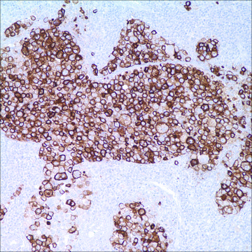
It has been reported that anti-SOX-2 antibody recognizes lung squamous cell carcinoma (LSCC). A recent study has demonstrated that extensive anti-SOX-2 staining was seen in over 90% of LSCC and largely paralleled p63 expression. Extensive anti-SOX-2 staining was seen in 21% of lung adenocarcinomas (LACA), including cases that were anti-p63-negative or only anti-p63 focally-positive. However, another recent study showed only 4.5% of LACA is positive for anti-SOX-2 expression. In a study by Sholl et al, 29% of LACA cases exhibited at least focal p63 expression. Combined p63 and SOX-2 expression was seen in 94% of LSCC and 12% of LACA with a statistically significant difference (P<0.0001) versus p63 alone. This study also showed that anti-CK5/6 had a good sensitivity but poor specificity for LSCC. Combined anti-CK5/6 and anti-p63 positivity was seen in 93% of LSCC and 24% of LACA. Anti-CK5/6+/anti-p63+/anti- SOX-2+ was detected in 93% of LSCC and only 9% of LACA. These results indicate that the sensitivity of anti-p63 is equally high but its specificity is similarly variable; it was seen at least focally in close to 30% of LACA. When used together, anti-p63+/anti-SOX-2+ applied to the same tumor cell population is >90% specific for LSCC. Anti-SOX-2 produced moderate-to-intense staining in all 50 cases of embryonal carcinoma components. The only other component that showed reactivity was the primitive neuroectodermal component in 11 of 14 (79%) of immature teratomas. In each of these positive staining foci, the staining varied from moderate-to-strong. Yolk sac tumor, seminoma, mature teratoma, choriocarcinoma, and IGCNU were uniformly negative, as were all the non-neoplastic parenchymal and stromal structures.

Quality


IVD

IVD

IVD

RUO

Linkage

SOX-2 Positive Control Slides, Product No. 371S, are available for immunohistochemistry (formalin-fixed, paraffin-embedded sections).

Physical form

Solution in Tris Buffer, pH 7.3-7.7, with 1% BSA and <0.1% Sodium Azide

Preparation Note

Download the IFU specific to your product lot and formatNote: This requires a keycode which can be found on your packaging or product label.

Other Notes

For Technical Service please contact: 800-665-7284 or email: service@cellmarque.com

Legal Information

Cell Marque is a registered trademark of Merck KGaA, Darmstadt, Germany

Not finding the right product?  

Try our Product Selector Tool.


Choose from one of the most recent versions:

Certificates of Analysis (COA)

Lot/Batch Number

Don't see the Right Version?

If you require a particular version, you can look up a specific certificate by the Lot or Batch number.

Already Own This Product?

Find documentation for the products that you have recently purchased in the Document Library.

Visit the Document Library

Our team of scientists has experience in all areas of research including Life Science, Material Science, Chemical Synthesis, Chromatography, Analytical and many others.

Contact Technical Service