Skip to Content
Merck

Skip To

HPA027287

Sigma-Aldrich

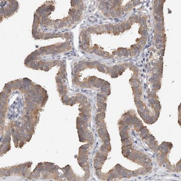
Anti-WDR47 antibody produced in rabbit

affinity isolated antibody, buffered aqueous glycerol solution, ab1

Synonym(s):

Anti-KIAA0893, Anti-WD repeat domain 47

Sign Into View Organizational & Contract Pricing

About This Item

UNSPSC Code:
12352203
Human Protein Atlas Number:
NACRES:
NA.43
Pricing and availability is not currently available.
Technical Service
Need help? Our team of experienced scientists is here for you.
Let Us Assist

biological source

rabbit

Quality Level

conjugate

unconjugated

antibody form

affinity isolated antibody

antibody product type

primary antibodies

clone

polyclonal

form

buffered aqueous glycerol solution

species reactivity

human

technique(s)

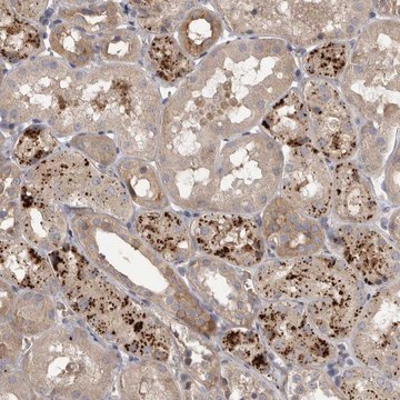
immunofluorescence: 0.25-2 μg/mL
immunohistochemistry: 1:20-1:50

immunogen sequence

LLYECCVEFCQSKATGEEITESEVLLGIDLLCGNGCDDLDLSLLSWLQNLPSSVFSCAFEQKMLNIHVDKLLKPTKAAYADLLTPLISKLSPYP

Compare Similar Items

View Full Comparison

Show Differences

1 of 4

This Item
HPA046719HPA026437HPA037376
Quality Level

100

Quality Level

100

Quality Level

100

Quality Level

100

conjugate

unconjugated

conjugate

unconjugated

conjugate

unconjugated

conjugate

unconjugated

antibody form

affinity isolated antibody

antibody form

affinity isolated antibody

antibody form

affinity isolated antibody

antibody form

affinity isolated antibody

biological source

rabbit

biological source

rabbit

biological source

rabbit

biological source

rabbit

Gene Information

human ... WDR47(22911)

Gene Information

human ... WDR44(54521)

Gene Information

human ... WDR77(79084)

Gene Information

human ... WDR37(22884)

UniProt accession no.

O94967

UniProt accession no.

Q5JSH3

UniProt accession no.

Q9BQA1

UniProt accession no.

Q9Y2I8

General description

WD repeat domain 47 (WDR47) is localized to cytoplasmic microtubules. The gene encoding this protein is localized on human chromosome 1p13.3.

Immunogen

WD repeat domain 47 recombinant protein epitope signature tag (PrEST)

Application

All Prestige Antibodies Powered by Atlas Antibodies are developed and validated by the Human Protein Atlas (HPA) project and as a result, are supported by the most extensive characterization in the industry.

The Human Protein Atlas project can be subdivided into three efforts: Human Tissue Atlas, Cancer Atlas, and Human Cell Atlas. The antibodies that have been generated in support of the Tissue and Cancer Atlas projects have been tested by immunohistochemistry against hundreds of normal and disease tissues and through the recent efforts of the Human Cell Atlas project, many have been characterized by immunofluorescence to map the human proteome not only at the tissue level but now at the subcellular level. These images and the collection of this vast data set can be viewed on the Human Protein Atlas (HPA) site by clicking on the Image Gallery link. We also provide Prestige Antibodies® protocols and other useful information.

Biochem/physiol Actions

WD repeat domain 47 (WDR47) interacts with ubiquitin carboxyl-terminal hydrolase 21 and microtubule-associated protein 8 (MAP8).

Features and Benefits

Prestige Antibodies® are highly characterized and extensively validated antibodies with the added benefit of all available characterization data for each target being accessible via the Human Protein Atlas portal linked just below the product name at the top of this page. The uniqueness and low cross-reactivity of the Prestige Antibodies® to other proteins are due to a thorough selection of antigen regions, affinity purification, and stringent selection. Prestige antigen controls are available for every corresponding Prestige Antibody and can be found in the linkage section.

Every Prestige Antibody is tested in the following ways:
  • IHC tissue array of 44 normal human tissues and 20 of the most common cancer type tissues.
  • Protein array of 364 human recombinant protein fragments.

Linkage

Corresponding Antigen APREST76551

Physical form

Solution in phosphate-buffered saline, pH 7.2, containing 40% glycerol and 0.02% sodium azide.

Legal Information

Prestige Antibodies is a registered trademark of Merck KGaA, Darmstadt, Germany

Disclaimer

Unless otherwise stated in our catalog or other company documentation accompanying the product(s), our products are intended for research use only and are not to be used for any other purpose, which includes but is not limited to, unauthorized commercial uses, in vitro diagnostic uses, ex vivo or in vivo therapeutic uses or any type of consumption or application to humans or animals.

Not finding the right product?  

Try our Product Selector Tool.

Storage Class Code

10 - Combustible liquids

WGK

WGK 1

Flash Point(F)

Not applicable

Flash Point(C)

Not applicable


Choose from one of the most recent versions:

Certificates of Analysis (COA)

Lot/Batch Number

Don't see the Right Version?

If you require a particular version, you can look up a specific certificate by the Lot or Batch number.

Already Own This Product?

Find documentation for the products that you have recently purchased in the Document Library.

Visit the Document Library

Anti-peptide monoclonal antibodies generated for immuno-MRM assays have a high probability of supporting Western blot and ELISA
Schoenherr RM, et al.
Molecular and Cellular Proteomics, mcp-O114 (2014)
Olivier Harismendy et al.
Genome biology, 12(12), R124-R124 (2011-12-22)
Ultra-deep targeted sequencing (UDT-Seq) can identify subclonal somatic mutations in tumor samples. Early assays' limited breadth and depth restrict their clinical utility. Here, we target 71 kb of mutational hotspots in 42 cancer genes. We present novel methods enhancing both
Root Gall Formation, Resting Spore Isolation and High Molecular Weight DNA Extraction of Plasmodiophora brassicae
Mehrabi S, et al.
Bio-protocol (2018)
Accurate variant detection across non-amplified and whole genome amplified DNA using targeted next generation sequencing
ElSharawy A, et al.
BMC Genomics, 13(1), 500-500 (2012)
Markus Frederik Schliffka et al.
eLife, 10 (2021-04-20)
During the first days of mammalian development, the embryo forms the blastocyst, the structure responsible for implanting the mammalian embryo. Consisting of an epithelium enveloping the pluripotent inner cell mass and a fluid-filled lumen, the blastocyst results from a series

Our team of scientists has experience in all areas of research including Life Science, Material Science, Chemical Synthesis, Chromatography, Analytical and many others.

Contact Technical Service