
05-1140
Anti-phospho-Focal Adhesion Kinase (Tyr397) Antibody, clone 18
clone 18, from mouse
Synonym(s):
FADK 1, PTK2 protein tyrosine kinase 2, Protein-tyrosine kinase 2, focal adhesion kinase 1
Sign Into View Organizational & Contract Pricing
Select a Size
All Photos(1)
Select a Size
Change View
About This Item
UNSPSC Code:
12352203
eCl@ss:
32160702
NACRES:
NA.41
Recommended Products


General description
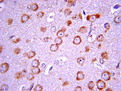
FAK plays a central role in cell spreading, differentiation, migration, cell death and acceleration of the G1 to S phase transition of the cell cycle. FAK regulation includes phosphorylation at multiple tyrosine and serine residues. Phosphorylation of tyrosine generally is associated with positive regulation and growth promotion, however, dephosphorylation at these sites occurs as cells enter mitosis (M-Phase of the cell cycle). In contrast, serine phosphorylation either remains high or is increased as cells enter mitosis and may play a role in focal adhesion disassembly. Tyrosine 397 is the autophosphorylation site of Focal Adhesion Kinase. The site binds Src family SH2 domains and the p85 subunit of PI3-Kinase.
Specificity
Reacts specifically with Focal Adhesion Kinase (FAK) when phosphorylated on Tyr397. FAK is a cytoplasmic tyrosine kinase that colocalizes with integrins in focal adhesions. This cellular localization is directed by a 125 amino acid sequence at the C-terminus called the "Focal Adhesion Targeting" sequence (FAT). The binding of extracellular matrix ligands to integrins triggers autophosphorylation at Tyr-397, and activation of FAK through phosphorylation of Tyr residues (Tyr-576 and Tyr577) in the kinase domain activation loop.
Immunogen
Epitope: Tyrosine 397
Generated from human FAK, a.a. 393-404, phosphorylated on Tyr397.
Application
Research Category
Cell Structure
Cell Structure
Research Sub Category
Cytoskeletal Signaling
Cytoskeletal Signaling
This Anti-phospho-Focal Adhesion Kinase (Tyr397) Antibody, clone 18 is validated for use in WB for the detection of phospho-Focal Adhesion Kinase (Tyr397).
Quality
Evaluated by Western Blot in LPS treated RAW 264 lysates.
Western Blot Analysis: 1:500 dilution of this lot detected phospho-FAK (Tyr397) on 10 ug of LPS treated RAW 264 lysates.
Western Blot Analysis: 1:500 dilution of this lot detected phospho-FAK (Tyr397) on 10 ug of LPS treated RAW 264 lysates.
Target description
125 kDa
Linkage
Replaces: 04-974
Physical form
Format: Purified
Protein A purified
Purified mouse monoclonal IgG1 in aqueous buffered solution containing 50% glycerol, BSA, and <0.09% sodium azide.
Storage and Stability
Stable for 1 year at -20ºC from date of receipt.
Note: Variability in freezer temperatures below -20°C may cause glycerol containing solutions to become frozen during storage.
Note: Variability in freezer temperatures below -20°C may cause glycerol containing solutions to become frozen during storage.
Analysis Note
Control
LPS treated RAW 264 lysates.
LPS treated RAW 264 lysates.
Other Notes
Concentration: Please refer to the Certificate of Analysis for the lot-specific concentration.
Disclaimer
Unless otherwise stated in our catalog or other company documentation accompanying the product(s), our products are intended for research use only and are not to be used for any other purpose, which includes but is not limited to, unauthorized commercial uses, in vitro diagnostic uses, ex vivo or in vivo therapeutic uses or any type of consumption or application to humans or animals.
Not finding the right product?
Try our Product Selector Tool.
Storage Class
10 - Combustible liquids
wgk_germany
WGK 2
flash_point_f
Not applicable
flash_point_c
Not applicable
Certificates of Analysis (COA)
Search for Certificates of Analysis (COA) by entering the products Lot/Batch Number. Lot and Batch Numbers can be found on a product’s label following the words ‘Lot’ or ‘Batch’.
Already Own This Product?
Find documentation for the products that you have recently purchased in the Document Library.
Our team of scientists has experience in all areas of research including Life Science, Material Science, Chemical Synthesis, Chromatography, Analytical and many others.
Contact Technical Service