
AB1922
Anti-Integrin β4 Antibody
serum, Chemicon®
Synonym(s):
CD104
About This Item
Recommended Products


biological source
rabbit
Quality Level
antibody form
serum
antibody product type
primary antibodies
clone
polyclonal
species reactivity
human, mouse, rat
manufacturer/tradename
Chemicon®
technique(s)
immunohistochemistry: suitable
immunoprecipitation (IP): suitable
NCBI accession no.
UniProt accession no.
shipped in
wet ice
target post-translational modification
unmodified
Gene Information
human ... ITGB4(3691)
mouse ... Itgb4(192897)
rat ... Itgb4(25724)
Specificity
Immunogen
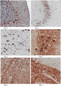
Application
Immunohistochemistry: 5 μg/mL. See protocol below.
Optimal working dilutions must be determined by the end user.
Suggested Protocol for Immunohistochemical:
1. Fix cryostat sections with a 1:1 chloroform-acetone mixture for 10 minutes at room temperature.
2. Air Dry.
3. Wash with 2 mL of 1x PBS.
4. Add 200 μL of first antibody (5 μg/mL) to each slide. Incubate for 1 hour at room temperature in a humidity chamber.
5. Wash with 5 mL of 1x PBS.
6. Add appropriately diluted anti-rabbit secondary antibody conjugate and incubate for 1 hour in a humidity chamber
7. Wash with 5 mL of 1x PBS.
8. Add 200 μL of development solution (containing DAB or other substrate depending on the secondary antibody) and incubate at room temperature for the necessary time to develop the dark color. Follow the reaction with a microscope, to establish the correct color intensity.
9. Block the reaction by washing with 2 mL of 1x PBS.
10. Counterstain with hematoxylin as desired.
Legal Information
Not finding the right product?
Try our Product Selector Tool.
Storage Class
10 - Combustible liquids
wgk_germany
WGK 1
Certificates of Analysis (COA)
Search for Certificates of Analysis (COA) by entering the products Lot/Batch Number. Lot and Batch Numbers can be found on a product’s label following the words ‘Lot’ or ‘Batch’.
Already Own This Product?
Find documentation for the products that you have recently purchased in the Document Library.
Our team of scientists has experience in all areas of research including Life Science, Material Science, Chemical Synthesis, Chromatography, Analytical and many others.
Contact Technical Service