
A5102
Anti-Aurora B antibody produced in rabbit
IgG fraction of antiserum, buffered aqueous solution
Synonym(s):
Anti-AIM-1, Anti-AIR-2 Kinase, Anti-AIRK-2
About This Item
Recommended Products


biological source
rabbit
Quality Level
conjugate
unconjugated
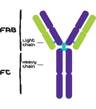
antibody form
IgG fraction of antiserum
antibody product type
primary antibodies
clone
polyclonal
form
buffered aqueous solution
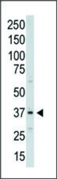
mol wt
antigen 41 kDa
species reactivity
rat, mouse, human
packaging
antibody small pack of 25 μL
1 of 4
This Item | A1231 | SAB1300093 | WH0009212M3 |
|---|---|---|---|
| Quality Level 200 | Quality Level 200 | Quality Level 200 | Quality Level 100 |
| conjugate unconjugated | conjugate unconjugated | conjugate unconjugated | conjugate unconjugated |
| biological source rabbit | biological source mouse | biological source rabbit | biological source mouse |
| antibody form IgG fraction of antiserum | antibody form purified immunoglobulin | antibody form IgG fraction of antiserum | antibody form purified immunoglobulin |
| UniProt accession no. | UniProt accession no. | UniProt accession no. | UniProt accession no. |
| species reactivity rat, mouse, human | species reactivity human, mouse | species reactivity human | species reactivity human |


General description
Specificity
Immunogen
Application
- in immunoblotting
- in immunoprecipitation
- in immunofluorescence
- in immunofluorescence staining
- in western blotting
Western Blotting (1 paper)
Biochem/physiol Actions
Target description
Physical form
Disclaimer
Not finding the right product?
Try our Product Selector Tool.
recommended
related product
Storage Class
12 - Non Combustible Liquids
wgk_germany
nwg
flash_point_f
Not applicable
flash_point_c
Not applicable
Choose from one of the most recent versions:
Already Own This Product?
Find documentation for the products that you have recently purchased in the Document Library.
Our team of scientists has experience in all areas of research including Life Science, Material Science, Chemical Synthesis, Chromatography, Analytical and many others.
Contact Technical Service