HPA001605
Anti-KRT18 antibody produced in rabbit

Prestige Antibodies® Powered by Atlas Antibodies, affinity isolated antibody, buffered aqueous glycerol solution
Synonym(s):
Anti-CK-18, Anti-Cell proliferation-inducing gene 46 protein, Anti-Cytokeratin 18, Anti-ENSG00000177083 antibody produced in rabbit, Anti-K18, Anti-Keratin 18
About This Item
Recommended Products
biological source
rabbit
Quality Level
antibody form
affinity isolated antibody
antibody product type
primary antibodies
clone
polyclonal
product line
Prestige Antibodies® Powered by Atlas Antibodies
form
buffered aqueous glycerol solution
species reactivity
human
enhanced validation
orthogonal RNAseq
Learn more about Antibody Enhanced Validation
technique(s)
immunoblotting: 0.04-0.4 μg/mL
immunofluorescence: 0.25-2 μg/mL
immunohistochemistry: 1:50-1:200
1 of 4
This Item | 27134 | 27150-U | 27172-U |
|---|---|---|---|
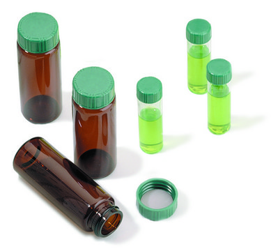
| feature closure type green melamine solid-top cap, closure type screw top vial | feature closure type green melamine solid-top cap, closure type screw top vial | feature closure type green melamine solid-top cap, closure type screw top vial | feature closure type green melamine solid-top cap, closure type screw top vial |
| volume 15 mL | volume 2 mL | volume 7 mL | volume 22 mL |
| fitting thread for 18-400 | fitting thread for 8-425 | fitting thread for 15-425 | fitting thread for 20-400 |
| material F217/PTFE cap liner, clear glass vial, PTFE liner, green melamine resin cap (solid-top) | material F217/PTFE cap liner, PTFE liner, clear glass vial (standard opening - 4.6 mm), green melamine resin cap (solid-top) | material F217/PTFE cap liner, PTFE liner, clear glass vial, green melamine resin cap (solid-top) | material F217/PTFE cap liner, PTFE liner, clear glass vial, green melamine resin cap (solid-top) |
| packaging pkg of 100 ea | packaging pkg of 100 ea | packaging pkg of 100 ea | packaging pkg of 100 ea |
| O.D. × H 21 mm × 70 mm | O.D. × H - | O.D. × H 17 mm × 60 mm | O.D. × H 23 mm × 85 mm |
Specificity
Immunogen
Application
The Human Protein Atlas project can be subdivided into three efforts: Human Tissue Atlas, Cancer Atlas, and Human Cell Atlas. The antibodies that have been generated in support of the Tissue and Cancer Atlas projects have been tested by immunohistochemistry against hundreds of normal and disease tissues and through the recent efforts of the Human Cell Atlas project, many have been characterized by immunofluorescence to map the human proteome not only at the tissue level but now at the subcellular level. These images and the collection of this vast data set can be viewed on the Human Protein Atlas (HPA) site by clicking on the Image Gallery link. We also provide Prestige Antibodies® protocols and other useful information.
Features and Benefits
Every Prestige Antibody is tested in the following ways:
- IHC tissue array of 44 normal human tissues and 20 of the most common cancer type tissues.
- Protein array of 364 human recombinant protein fragments.
Linkage
Physical form
Legal Information
Not finding the right product?
Try our Product Selector Tool.
Storage Class
10 - Combustible liquids
wgk_germany
WGK 1
flash_point_f
Not applicable
flash_point_c
Not applicable
Choose from one of the most recent versions:
Certificates of Analysis (COA)
Don't see the Right Version?
If you require a particular version, you can look up a specific certificate by the Lot or Batch number.
Already Own This Product?
Find documentation for the products that you have recently purchased in the Document Library.
Our team of scientists has experience in all areas of research including Life Science, Material Science, Chemical Synthesis, Chromatography, Analytical and many others.
Contact Technical Service


