M4528
Monoclonal Anti-MAP1b antibody produced in mouse
clone AA6, ascites fluid
Synonym(s):
Anti-MAP1.2, Anti-MAP1X, Anti-MAP5
Sign Into View Organizational & Contract Pricing
Select a Size
All Photos(2)
Select a Size
Change View
About This Item
Recommended Products
biological source
mouse
Quality Level
conjugate
unconjugated
antibody form
ascites fluid
antibody product type
primary antibodies
clone
AA6, monoclonal
contains
15 mM sodium azide
species reactivity
human, feline, bovine, hamster, chicken, mouse, rat
technique(s)
microarray: suitable
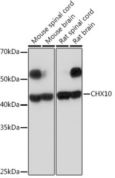
western blot: 1:500 using a fresh total rat brain extract or an enriched microtubule protein preparation
isotype
IgG1
General description
MAP1b is a microtubule associated protein that interacts with actin filaments. MAP1b regulates microtubule stabilization and the development of axons. It may also act as a scaffolding protein Addition of antibody to microtubule proteins before polymerization in immunoassays results in abnormally short (but otherwise morphologically normal) microtubules. Immunohistochemical staining of brain tissue with the product shows selective labeling of dendritic trees throughout the brain. Monoclonal Anti-MAP1b does not react with tubulin of other microtubule associated proteins.
Monoclonal Anti-MAP1b (MAP5) (mouse IgG1 isotype) is derived from the hybridoma produced by the fusion of mouse myeloma cells and splenocytes from an immunized mouse. Microtubule-associated protein (MAP1B) is the major microtubule associated protein in developing brain which changes its expression during development. In the new born rat brain, it is a major component of microtubules but in the adult its level is ten-fold lower.
Specificity
Monoclonal Anti-MAP1b antibody is specific for MAP1b in humans, mice, rats, bovines, cats, hamsters and chickens. The antibody does not cross-react with other MAPs or tubulin. Addition of the antibody to microtubule proteins before polymerization results in abnormally short (but otherwise morphologically normal) microtubules. In immunohistochemical staining of brain tissue, the antibody shows selective labeling of neurons with stronger staining of axons, dendrites and cell bodies.
Immunogen
rat brain microtubule-associated proteins (MAPs)
Application
Monoclonal Anti-MAP1b antibody produced in mouse has been used in:
- immunohistochemistry
- immunostaining
- fluorescence microscopy
Biochem/physiol Actions
Microtubule-associated protein (MAP1B) is the first MAP to appear in growing axons during development as it is present from the first emergence of the nascent axon from the cell body. Monoclonal Anti-MAP1b may be used to study MAP expression and cytological localization both in tissues and cell lines, under different developmental and environmental circumstances.
Disclaimer
Unless otherwise stated in our catalog or other company documentation accompanying the product(s), our products are intended for research use only and are not to be used for any other purpose, which includes but is not limited to, unauthorized commercial uses, in vitro diagnostic uses, ex vivo or in vivo therapeutic uses or any type of consumption or application to humans or animals.
Not finding the right product?
Try our Product Selector Tool.
Storage Class
10 - Combustible liquids
wgk_germany
WGK 2
flash_point_f
Not applicable
flash_point_c
Not applicable
Choose from one of the most recent versions:
Already Own This Product?
Find documentation for the products that you have recently purchased in the Document Library.
A Matus et al.
Journal of neurochemistry, 49(3), 714-720 (1987-09-01)
The influence on microtubule assembly in vitro of monoclonal antibodies against microtubule-associated proteins (MAPs) was studied. Light scattering was used for measuring net polymer formation and electron microscopy for determining the influence of antibodies on microtubule morphology. Control experiments showed
Human mesenchymal precursor cells (Stro-1+) from spinal cord injury patients improve functional recovery and tissue sparing in an acute spinal cord injury rat model
Hodgetts SI, et al.
Cell Transplantation, 22(3), 393-412 (2013)
Association of microtubule-associated protein (MAP1B) with growing axons in cultured hippocampal neurons
Fischer I and Romano CG
Molecular and Cellular Neurosciences, 2(1), 39-51 (1991)
Rebecca L Anderson et al.
The Journal of comparative neurology, 447(3), 218-233 (2002-05-02)
Visceromotor neurons in mammalian prevertebral sympathetic ganglia receive convergent synaptic inputs from spinal preganglionic neurons and peripheral intestinofugal neurons projecting from the enteric plexuses. Vasomotor neurons in the same ganglia receive only preganglionic inputs. How this pathway-specific pattern of connectivity
J B Denny
Journal of neurocytology, 20(8), 627-636 (1991-08-01)
A monoclonal antibody was used to determine both the expression of the microtubule-associated protein MAP5 in cultured foetal rat hippocampal neurons as a function of culture age and the cellular distribution of the protein. When cultures at days 2 and
Our team of scientists has experience in all areas of research including Life Science, Material Science, Chemical Synthesis, Chromatography, Analytical and many others.
Contact Technical Service