Skip to Content
MilliporeSigma

Skip To

SAB4200678

Sigma-Aldrich

Anti-Collagen Type I antibody, Mouse monoclonal

clone COL-1, purified from hybridoma cell culture

Synonym(s):

Monoclonal Anti-Collagen Type I antibody produced in mouse, OI4, alpha 1, alpha 2, collagen, type I

Sign Into View Organizational & Contract Pricing

Select a Size

100 μL
$635.00

$635.00


Please contact Customer Service for Availability

Request a Bulk Order

Select a Size

Change View
100 μL
$635.00

About This Item

UNSPSC Code:
51111800
NACRES:
NA.41

$635.00


Please contact Customer Service for Availability

Request a Bulk Order
Technical Service
Need help? Our team of experienced scientists is here for you.
Let Us Assist

biological source

mouse

Quality Level

antibody form

purified immunoglobulin

antibody product type

primary antibodies

clone

COL-1, monoclonal

form

buffered aqueous solution

species reactivity

pig, rat, human, deer, bovine

concentration

~1 mg/mL

technique(s)

ELISA: suitable
dot blot: suitable
immunoblotting: 1-2 μg/mL using recombinant human collagen, expressed in Nicotiana tabacum
immunohistochemistry: 3.5-7 μg/mL using frozen sections of human tonsil, human tongue or pig tongue

isotype

IgG1

Compare Similar Items

View Full Comparison

Show Differences

1 of 4

This Item
SML0022SML0886SML2465
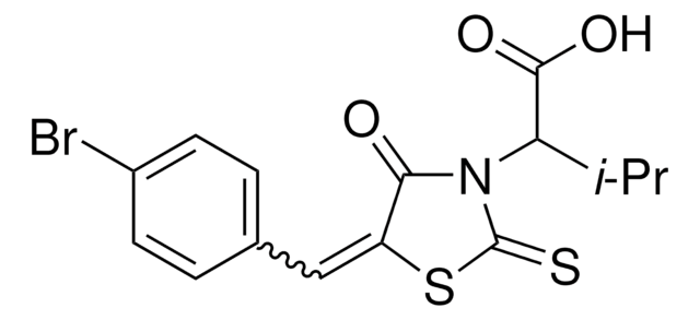
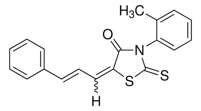
BH3I-1 ≥97% (HPLC), powder, yellow

B8809

BH3I-1

IMB-10 ≥98% (HPLC)

SML0022

IMB-10

CY-09 ≥98% (HPLC)

SML2465

CY-09

assay

≥97% (HPLC)

assay

≥98% (HPLC)

assay

≥98% (HPLC)

assay

≥98% (HPLC)

form

powder, yellow

form

powder

form

powder

form

powder

Quality Level

100

Quality Level

100

Quality Level

100

Quality Level

-

storage temp.

2-8°C

storage temp.

2-8°C

storage temp.

2-8°C

storage temp.

2-8°C

solubility

DMSO: soluble >10 mg/mL, H2O: insoluble

solubility

DMSO: ≥25 mg/mL

solubility

DMSO: 3 mg/mL, clear (warmed)

solubility

DMSO: 2 mg/mL, clear

General description

Collagen type Iα1 (COL1A1) is encoded by the gene that is located on human chromosome 17q21.33. It is the most abundant extracellular matrix (ECM) protein in humans. Type 1 collagen is the major structural protein of bone, tendon, skin and cornea. The encoded protein is a heterotrimer consisting of two α1-chains and one α2-chain.
Collagens are present in the extracellular matrix and contains a chains which can differ in length.

Immunogen

bovine skin collagen type I

Application

Monoclonal Anti-Collagen Type I antibody produced in mouse has been used in immunohistochemical analysis and in vitro invasion assays.

Biochem/physiol Actions

Collagen, type I (COL1A1) is an important component of the connective tissue matrix. It plays an important role in the growth and maintenance of organ and tissue integrity. This protein also participates in the process of tissue repair. COL1A1 is implicated in fibrosis.
Collagens are involved in structural roles. Additionally, they associate with various receptors and control cell proliferation, migration as well as differentiation.

Physical form

Solution in 0.01 M phosphate buffered saline pH 7.4, containing 15 mM sodium azide.

Disclaimer

Unless otherwise stated in our catalog or other company documentation accompanying the product(s), our products are intended for research use only and are not to be used for any other purpose, which includes but is not limited to, unauthorized commercial uses, in vitro diagnostic uses, ex vivo or in vivo therapeutic uses or any type of consumption or application to humans or animals.

Not finding the right product?  

Try our Product Selector Tool.

Storage Class Code

12 - Non Combustible Liquids

WGK

WGK 1

Flash Point(F)

Not applicable

Flash Point(C)

Not applicable


  • Choose from one of the most recent versions:

    Certificates of Analysis (COA)

    Lot/Batch Number

    Don't see the Right Version?

    If you require a particular version, you can look up a specific certificate by the Lot or Batch number.

    Already Own This Product?

    Find documentation for the products that you have recently purchased in the Document Library.

    Visit the Document Library

    Expression of G protein-coupled receptor 30 in the spinal somatosensory system.
    Keiko Takanami,Hirotaka Sakamoto,Ken-Ichi Matsuda,Koji Hosokawa,Mayumi Nishi et al.
    Brain Research null
    Andrea M Sartori et al.
    Experimental neurology, 348, 113937-113937 (2021-11-27)
    Neurogenic lower urinary tract dysfunction typically develops after spinal cord injury. We investigated the time course and the anatomical changes in the spinal cord that may be causing lower urinary tract symptoms following injury. Rats were implanted with a bladder
    Maria N Marangoni et al.
    Cardiovascular diabetology, 13, 11-11 (2014-01-15)
    Cardiomyopathy and distal symmetrical polyneuropathy (DSPN), including sensory and autonomic dysfunction, often co-occur in diabetic mellitus (DM) patients. However, the temporal relationship and progression between these two complications has not been investigated. Using a streptozotocin DM animal model that develops
    Aline Dionizio et al.
    Chemosphere, 273, 129607-129607 (2021-01-29)
    Gastrointestinal signs and symptoms are the first signs of toxicity due to exposure to fluoride (F). This suggests the possibility that lower levels of subchronic F exposure may affect the gut. The aim of this study was to evaluate changes
    Michał Bulc et al.
    Journal of diabetes research, 2018, 4735659-4735659 (2018-08-25)
    One of the most frequently reported disorders associated with diabetes is gastrointestinal (GI) disturbance. Although pathogenesis of these complications is multifactorial, the complicity of the enteric nervous system (ENS) in this respect has significant importance. Therefore, this paper analysed changes

    Our team of scientists has experience in all areas of research including Life Science, Material Science, Chemical Synthesis, Chromatography, Analytical and many others.

    Contact Technical Service