
추천 제품


생물학적 소스
mouse
Quality Level
항체 형태
purified immunoglobulin
항체 생산 유형
primary antibodies
클론
4-10-C9, monoclonal
종 반응성
mouse
기술
flow cytometry: suitable
immunocytochemistry: suitable
동형
IgG2aκ
NCBI 수납 번호
UniProt 수납 번호
배송 상태
wet ice
타겟 번역 후 변형
unmodified
유전자 정보
mouse ... Lag3(16768)
일반 설명
Lymphocyte activation gene 3 protein (UniProt Q61790; also known as CD223, LAG-3) is encoded by the Lag3 gene (Gene ID 16768) in murine species. LAG-3 is an inhibitory T-cell surface molecule that modulates T-cell activation and homeostasis. LAG-3 co-localizes with CD8 and CD4 upon TCR engagement and alters TCR signaling. LAG-3 suppresses homeostasis proliferation (HP) of both lymphocytes and some dendritic cell populations in vivo, and enhanced HP is observed in mice deficient in LAG-3. LAG-3 signaling is shown to negatively regulate STAT5 phosphorylation in activated T cells, and no enhancement of HP was seen upon LAG-3 blockade in mice deficient in both STAT5a and STAT5b. Murine LAG-3 is produced with a signal peptide sequence (a.a. 1-22), the removal of which yields the mature protein with a large extracellular region (a.a. 23-442), followed by a transmembrane segment (a.a. 443-463) and a cytoplasmic tail (a.a. 464-521). The extracellular portion contains a V-type Ig-like domain (a.a. 37-163), followed by three C2-type Ig-like domains (a.a. 165-246, 258-341, and 345-412) and elven tandem repeats of 2-amino acid E-X seqeunce at its C-terminal end (a.a. 493-518).
특이성
Clone 4-10-C9 immunostained surface LAG-3 on activated CD4+ T cells by targeting an extracellular epitope within the third and fourth Ig-like domains (D3/D4 domains; second and third C2-type Ig-like domains). Surface LAG-3 degradation by pronase treatment abolished cell surface staining (Woo, S.R., et al. (2010). Eur. J. Immunol. 40(6):1768-1777).
면역원
Epitope: Within D3/D4 domains.
Murine LAG-3-expressing mouse T-cell hybridoma.
애플리케이션
Flow Cytometry Analysis: 2.5 µg/mL from a representative lot detected surface LAG-3 immunoreactivity among the CD4+ and CD8+ populations of wild-type, but not Lag3-knockout, mouse splenocytes activated in vitro via CD3 cross-linking (Courtesy of Dario A. Vignali, Ph.D., University of Pittsburgh, PA, U.S.A.).
Flow Cytometry Analysis: A representative lot was fluorescently conjugated and detected an increased number of LAG-3-positive cells within the CD4+ and CD8+ populations of infiltrating lymphocytes (TILs) in tumors developed in mice exografted with murine B16 melanoma, MC38 colon adenocarcinoma, or Sa1N fibrosarcoma cells (Woo, S.R., et al. (2012). Cancer Res. 72(4): 917–927).
Flow Cytometry Analysis: A representative lot, pre-conjugated with Alexa Fluor™ 647, detected both surface and intracellular LAG-3 by immunofluorescent staining of non-permeabilized and permeabilized primary murine CD4+ T cells activated in vitro via CD3 & CD28 cross-linking by immobilized antibodies. Pronase treatment of cells prior to permeabilization abolished cell surface staining (Woo, S.R., et al. (2010). Eur. J. Immunol. 40(6):1768-1777).
Flow Cytometry Analysis: A representative lot, pre-conjugated with Alexa Fluor 647, detected a time-dependent recovery of cell surface LAG-3 immunoreactivity on activated murine CD4+ T cells after initial surface LAG-3 degradation by pronase treatment. Protein synthesis inhibitor cycloheximide (Cat. No. 239764) or protein transport inhibitor Brefeldin A (Cat. No. 203729) treatment partially blocked the recovery (Woo, S.R., et al. (2010). Eur. J. Immunol. 40(6):1768-1777).
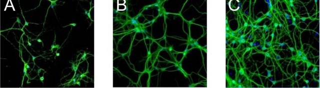
Immunocytochemistry Analysis: A representative lot detected both surface and intracellular LAG-3 by fluorescent immunocytochemistry staining of non-permeabilized and permeabilized primary murine CD4+ T cells activated in vitro via CD3 & CD28 cross-linking by immobilized antibodies. Pronase treatment of cells prior to permeabilization abolished cell surface staining (Woo, S.R., et al. (2010). Eur. J. Immunol. 40(6):1768-1777).
Immunocytochemistry Analysis: A representative lot detected intracellular LAG-3 immunoreactivity co-localized with those of the early and recycling endosome marker EEA1, as well as endosomal markers Rab11b and Rab27a by fluorescent immunocytochemistry staining of activated murine CD4+ T cells following pronase treatment and permeabilization (Woo, S.R., et al. (2010). Eur. J. Immunol. 40(6):1768-1777).
Flow Cytometry Analysis: A representative lot was fluorescently conjugated and detected an increased number of LAG-3-positive cells within the CD4+ and CD8+ populations of infiltrating lymphocytes (TILs) in tumors developed in mice exografted with murine B16 melanoma, MC38 colon adenocarcinoma, or Sa1N fibrosarcoma cells (Woo, S.R., et al. (2012). Cancer Res. 72(4): 917–927).
Flow Cytometry Analysis: A representative lot, pre-conjugated with Alexa Fluor™ 647, detected both surface and intracellular LAG-3 by immunofluorescent staining of non-permeabilized and permeabilized primary murine CD4+ T cells activated in vitro via CD3 & CD28 cross-linking by immobilized antibodies. Pronase treatment of cells prior to permeabilization abolished cell surface staining (Woo, S.R., et al. (2010). Eur. J. Immunol. 40(6):1768-1777).
Flow Cytometry Analysis: A representative lot, pre-conjugated with Alexa Fluor 647, detected a time-dependent recovery of cell surface LAG-3 immunoreactivity on activated murine CD4+ T cells after initial surface LAG-3 degradation by pronase treatment. Protein synthesis inhibitor cycloheximide (Cat. No. 239764) or protein transport inhibitor Brefeldin A (Cat. No. 203729) treatment partially blocked the recovery (Woo, S.R., et al. (2010). Eur. J. Immunol. 40(6):1768-1777).
Immunocytochemistry Analysis: A representative lot detected both surface and intracellular LAG-3 by fluorescent immunocytochemistry staining of non-permeabilized and permeabilized primary murine CD4+ T cells activated in vitro via CD3 & CD28 cross-linking by immobilized antibodies. Pronase treatment of cells prior to permeabilization abolished cell surface staining (Woo, S.R., et al. (2010). Eur. J. Immunol. 40(6):1768-1777).
Immunocytochemistry Analysis: A representative lot detected intracellular LAG-3 immunoreactivity co-localized with those of the early and recycling endosome marker EEA1, as well as endosomal markers Rab11b and Rab27a by fluorescent immunocytochemistry staining of activated murine CD4+ T cells following pronase treatment and permeabilization (Woo, S.R., et al. (2010). Eur. J. Immunol. 40(6):1768-1777).
This Anti-LAG3 Antibody, clone 4-10-C9 is validated for use in Flow Cytometry, Immunocytochemistry for the detection of LAG3.
품질
Evaluated by Flow Cytometry in mouse splenocytes.
Flow Cytometry Analysis: 1 µg/mL of this antibody detected an induction of LAG-3-positive population in isolated mouse splenocytes following a 3-day 2 µg/mL Concanavalin A (Con A) stimulation.
Flow Cytometry Analysis: 1 µg/mL of this antibody detected an induction of LAG-3-positive population in isolated mouse splenocytes following a 3-day 2 µg/mL Concanavalin A (Con A) stimulation.
표적 설명
54.51/56.98 kDa (mature/pro-form) calculated.
물리적 형태
Format: Purified
법적 정보
ALEXA FLUOR is a trademark of Life Technologies
적합한 제품을 찾을 수 없으신가요?
당사의 제품 선택기 도구.을(를) 시도해 보세요.
Storage Class Code
12 - Non Combustible Liquids
WGK
WGK 1
시험 성적서(COA)
제품의 로트/배치 번호를 입력하여 시험 성적서(COA)을 검색하십시오. 로트 및 배치 번호는 제품 라벨에 있는 ‘로트’ 또는 ‘배치’라는 용어 뒤에서 찾을 수 있습니다.
자사의 과학자팀은 생명 과학, 재료 과학, 화학 합성, 크로마토그래피, 분석 및 기타 많은 영역을 포함한 모든 과학 분야에 경험이 있습니다..
고객지원팀으로 연락바랍니다.