
MABF291
Anti-S100A8/S100A9 Antibody, clone 5.5
clone 5.5, from mouse
Synonym(s):
Protein S100-A9, Calgranulin-B, Calprotectin L1H subunit, Leukocyte L1 complex heavy chain, Migration inhibitory factor-related protein 14, S100 calcium-binding protein A9, AMRP-14, p14, Protein S100-A8, Calgranulin-A, Calprotectin L1L subunit, Cystic fi
About This Item
Recommended Products


biological source
mouse
Quality Level
antibody form
purified immunoglobulin
antibody product type
primary antibodies
clone
5.5, monoclonal
species reactivity
human
technique(s)
ELISA: suitable
flow cytometry: suitable
immunohistochemistry: suitable
immunoprecipitation (IP): suitable
western blot: suitable
isotype
IgG1κ
1 of 4
This Item | MABF290 | MABC653 | MAB1789 |
|---|---|---|---|
| clone 5.5, monoclonal | clone 2A5, monoclonal | clone 9D5.1, monoclonal | clone AHN-17, monoclonal |
| antibody form purified immunoglobulin | antibody form purified antibody | antibody form purified immunoglobulin | antibody form purified immunoglobulin |
| Gene Information human ... S100A8(6279), S100A9(6280) | Gene Information human ... S100A9(6280) | Gene Information human ... S100A4(6275) | Gene Information human ... S100A8(6279) |
| biological source mouse | biological source rat | biological source mouse | biological source mouse |
| species reactivity human | species reactivity human, mouse | species reactivity human, mouse | species reactivity human |
| isotype IgG1κ | isotype IgG1 | isotype IgG1κ | isotype IgG1 |

General description
Specificity
Immunogen
Application
Inflammation & Immunology
Immunological Signaling
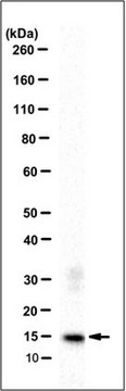
Western Blotting Analysis: A representative lot of this antibody was used to detect S100A8/S100A9 in neutrophil extracts (Hogg et al., 1989).
Immunoprecipitation Analysis: A representative lot of this antibody was used to detect S100A8/S100A9 in Human monocyte and neutrophil lysate (Edgeworth, J., et al., (1991) JBC. 266(12):7706-7713).
Immunoprecipitation Analysis: A representative lot of this antibody was used to detect S100A8/S100A9 in MRP-8 and TL-14 mutant lysate (Hessian P.A., et al., (2001) Eur. J. Biochem. 268:353-363).
Immunohistochemistry Analysis: A representative lot of this antibody was used to detect S100A8/S100A9 in Human Bronchus tissue (Hogg, N., et al., (1989) Eur. J. Immunol. 19:1053-1061).
Immunohistochemistry Analysis: A representative lot of this antibody was used to detect S100A8/S100A9 in spleen and thymus tissue (Hogg, N., et al., (1989) Eur. J. Immunol. 19:1053-1061).
ELISA: A representative lot of this antibody was used to detect S100A8/S100A9 in ELISA (Ryckman, C., et al., (2003) Arthritis & Rheumatism. 48(8):2310-2320).
Quality
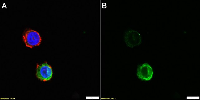
Flow Cytometry Analysis: A 1:80 dilution (0.25 µg) of this antibody detected S100A8 and/or S100A9 in 1x10E6 PBMCs.
Target description
Physical form
Storage and Stability
Other Notes
Disclaimer
Not finding the right product?
Try our Product Selector Tool.
Storage Class
12 - Non Combustible Liquids
wgk_germany
WGK 1
flash_point_f
Not applicable
flash_point_c
Not applicable
Certificates of Analysis (COA)
Search for Certificates of Analysis (COA) by entering the products Lot/Batch Number. Lot and Batch Numbers can be found on a product’s label following the words ‘Lot’ or ‘Batch’.
Already Own This Product?
Find documentation for the products that you have recently purchased in the Document Library.
Our team of scientists has experience in all areas of research including Life Science, Material Science, Chemical Synthesis, Chromatography, Analytical and many others.
Contact Technical Service